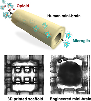
Researchers are 3D printing mini-brains to study the effects of multiple treatments.
Bioengineers at the Luddy School of Informatics, Computing, and Engineering are teaming up with neuroscientists from the Gill Center for Biomolecular Science at IU and the Stark Neurosciences Research Institute at the IU School of Medicine to create mini brains that can recapitulate human brain activity and function for studying neurological diseases such as drug abuse and neurodegeneration.
In “Tubular human brain organoids to model microglia-mediated neuroinflammation”, a study published in the journal Lab on a Chip, Postdoctoral Fellow of Intelligent Systems Engineering Zheng Ao and coworkers detail how the use of 3D-printed hollow mesh scaffolds to support brain tissue cultures derived from human pluripotent stem cells are showing promise to model key features of the developing human brain.
“My lab has been engineering human mini-brains or human brain organoids, human stem cell-derived 3D cultures to better understand brain development, function, and neural disorders,” said Feng Guo, assistant professor of intelligent systems engineering and the principal investigator of the Intelligent BioMedical Systems Lab at IU. “We realized that the current organoids can be flawed as they lack reproducibility and key innate immune components. Thus, based on our expertise in biomedical devices, we improved this model by incorporating 3D-printed scaffolds to enhance the reliability of the organoid culture as well as promote the integration of the brain's innate immune cell type, microglia into the culture.”
Organoids are three-dimensional tissue cultures that are derived from stem cells and can replicate the complexity of an organ at a smaller scale. By introducing rocking flows through the tubular organoid, researchers created a device that improves survival and oxygen levels of cultures, while also incorporating immune cells to model neuro-immune interactions. They then used these improved brain organoids to model the microglia-mediated neuroinflammation that occurs after exposure to an opioid receptor agonist.
“We wanted to better understand how the human brain responds to opioids and other substances,” Gill Chair and Distinguished Professor of Psychological and Brain Sciences Ken Mackie said. “We also demonstrated that the mini-brain model could mimic some of the key phenomena of human prenatal exposure to cannabis, so it provides us with a model to study the effects of abused drug on the developing human brain.”
“Microglia-mediated neuroinflammation plays a critical role in Alzheimer’s disease.” P. Michael Conneally Professor of Medical and Molecular Genetics Jungsu Kim said. “The potential impact of the mini-brain model is to study how patient genetics could impact neuroinflammation, as well as discover novel treatments by growing mini-brains from patient-derived stem cells.”
The model will be also used to study how developmental disorders, neurodegenerative diseases, cancer, and autoimmune disorders impact brain components and brain function.
“The use of 3D printer technology in the biomedical realm creates so many possibilities for the discovery of treatments and improvements in healthcare,” said Kay Connelly, associate dean for research at the Luddy School. “The work being conducted by Feng and his colleagues continues to open pathways and is a fantastic example of novel thinking and technology making a real-world impact.”
Other IU researchers contributing to the project were Hongwei Cai, graduate student; Zhuhao Wu, graduate student; Sunghwa Song, graduate student; all in intelligent systems engineering. Also involved with the project were postdoctoral fellows Hande Karahan and Byungwook Kim in the Stark Neurosciences Research Institute, and Hui-Chen Lu, Gill Chair and Professor of Psychological and Brain Sciences in IU’s Gill Center for Biomolecular Science.

